
At the office of Vaccaro Aesthetic and Family Dentistry, we integrate advanced imaging into everyday care so patients receive more accurate diagnoses and more predictable treatment results. Cone-beam computed tomography (CBCT) has become an important tool for modern dental practices because it reveals three-dimensional information that conventional X-rays cannot. This additional detail helps clinicians plan procedures with greater confidence while keeping patient comfort and safety front of mind.
CBCT captures high-resolution 3D images of teeth, bone, nerves, and sinuses in a single, quick scan. Those images give clinicians a fuller view of the oral and maxillofacial anatomy, which supports decision-making across restorative, surgical, and diagnostic treatments. Below are five focused sections that explain how CBCT works, why it matters, what patients can expect, and how we apply the technology to improve outcomes.
Traditional two-dimensional X-rays provide valuable information, but they compress complex anatomy into a flat image. CBCT removes that limitation by rendering volumetric data that clinicians can examine from multiple angles. That extra dimension makes it easier to detect hidden problems, appreciate the relationships between structures, and identify conditions that might otherwise be missed or underestimated.
For example, small variations in root anatomy, the proximity of nerves to an implant site, or the extent of a lesion are often clearer on a CBCT scan than on a periapical film or panoramic X-ray. This clarity reduces guesswork and helps clinicians anticipate and avoid potential complications before a procedure begins. In short, CBCT elevates diagnostic accuracy in ways that directly influence treatment planning.
Importantly, three-dimensional imaging is not used indiscriminately. We apply CBCT selectively when the additional information will change the course of care—such as prior to implant placement, for complex endodontic evaluations, or when assessing trauma and pathology. Using the right test for the right patient ensures that each scan adds clinical value.
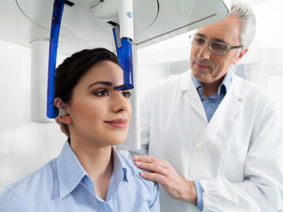
Cone-beam computed tomography acquires a series of low-dose X-ray images while the scanner rotates around the patient’s head. These images are reconstructed into a single, three-dimensional dataset that can be sliced and viewed in multiple planes. The result is a detailed map of bone, teeth, and surrounding structures without the need for invasive exploration.
Compared with medical CT scanners, dental CBCT units are designed specifically for head and neck imaging and typically deliver much lower radiation exposure. Scans are localized, meaning only the area of interest is captured rather than the entire mid-face or skull. Modern equipment and software also let clinicians choose appropriately sized fields of view to further minimize exposure while still obtaining diagnostic detail.
The scan itself is brief—often completed in less than a minute—and the images are available almost immediately for review. Because the procedure is fast and noninvasive, it is well suited to a wide range of patients, including those undergoing comprehensive treatment planning or evaluation for unexplained symptoms.
One of the most common uses of CBCT in dentistry is implant planning. Three-dimensional imaging allows the clinician to measure bone height and width, locate vital structures such as the inferior alveolar nerve and maxillary sinuses, and choose optimal implant size and angulation. This level of preoperative insight supports precise placement and reduces intraoperative surprises.
CBCT is also valuable in endodontics for detecting root fractures, locating accessory canals, and assessing complex root morphology that can influence the success of root canal therapy. In cases of dental trauma, CBCT helps evaluate fractures, tooth displacement, and bone involvement with a clarity that guides immediate and follow-up care.
Beyond restorative and endodontic care, CBCT plays a role in orthodontics, airway assessment, and temporomandibular joint (TMJ) analysis. It can reveal impacted teeth, assess airway volume for sleep-related breathing concerns, and display joint anatomy to help diagnose degenerative changes. Because the scan captures the full context of the jaws and adjacent structures, it supports multidisciplinary planning when multiple specialists are involved.
Finally, CBCT can assist in identifying pathology such as cysts or other lesions that require further investigation. When a suspicious area is discovered, three-dimensional imaging helps delineate its size, extent, and relationship to nearby teeth and anatomical landmarks—information that is essential for safe, effective management.
Preparation for a CBCT scan is minimal. Patients are usually asked to remove jewelry, eyeglasses, or removable dental appliances from the head and neck area. The scan is performed while the patient stands or sits in the machine with their head stabilized for a short period. Because motion degrades image quality, remaining still for the duration of the rotation is important but rarely difficult for most people.
The procedure itself is quick and comfortable. The scanner rotates once around the head and captures all necessary data in a single pass; many scans take less than a minute to complete. There is no need for injections or contrast agents for routine dental imaging, and the team monitors each step to ensure patient comfort and positioning are correct.
After the scan, images are reviewed by the clinical team and reconstructed into views that can be examined in detail. The dentist will interpret the findings in the context of your overall oral health and treatment goals, explaining what the images reveal and how that information informs recommended next steps.
Reading a CBCT dataset requires training and experience. Our clinicians combine their knowledge of dental anatomy with the scan’s three-dimensional information to form a comprehensive assessment. In many cases, CBCT findings are integrated with clinical exams, intraoral photographs, and conventional X-rays to produce a complete diagnostic picture.
Safety is a priority when using any imaging modality. We follow established guidelines to justify and optimize each scan, selecting the smallest field of view and appropriate settings to answer the clinical question while minimizing radiation dose. When imaging is not expected to change management, alternative methods are considered instead.
CBCT is best thought of as a complementary tool rather than a replacement for other diagnostic resources. By combining CBCT with clinical evaluation and other imaging modalities, clinicians can plan treatments with greater precision, communicate findings more clearly to patients, and coordinate care across specialties when needed.
At every stage, the focus remains on delivering thoughtful, patient-centered care that balances diagnostic benefit with safety and comfort.
Summary: CBCT adds a valuable layer of diagnostic detail that supports safer, more predictable dental care. If you have questions about whether three-dimensional imaging is appropriate for your situation, please contact us to learn more and discuss how CBCT could be used as part of your treatment plan.


Cone-beam computed tomography, commonly called CBCT, is an imaging technique that produces three-dimensional views of the teeth, jaws, and surrounding structures. It acquires a series of low-dose X-ray images while rotating around the head and reconstructs them into a volumetric dataset. Clinicians can view that dataset in multiple planes to examine anatomy with greater precision than conventional radiographs.
CBCT captures high-resolution detail of bone, tooth roots, nerves, and sinuses in a single scan and is especially useful when two-dimensional films are insufficient. Scans are typically brief and do not require injections or contrast agents, making the procedure comfortable for most patients. At Vaccaro Aesthetic and Family Dentistry we apply CBCT selectively to answer specific diagnostic questions and to support predictable treatment planning.
Traditional dental X-rays, such as periapical or panoramic films, provide flattened two-dimensional images that can obscure depth and spatial relationships. In contrast, CBCT renders volumetric data so clinicians can rotate, slice, and measure anatomy in three dimensions. This capability reduces ambiguity when assessing complex root systems, bony defects, or the relationship between planned implants and adjacent structures.
Because CBCT can be tailored with different fields of view and resolution settings, clinicians choose the most appropriate imaging modality for each case rather than using CBCT universally. Two-dimensional X-rays remain valuable for routine screening and follow-up when the added information from three-dimensional imaging would not change management. Selecting the right test helps balance diagnostic benefit with patient exposure and convenience.
CBCT units used in dental practices are designed for head and neck imaging and generally deliver much lower radiation than full medical CT scans while still providing three-dimensional detail. Clinicians follow principles such as ALARA—'as low as reasonably achievable'—to limit dose by selecting the smallest field of view and appropriate exposure settings. Modern equipment and software allow for dose optimization so that each scan is justified by a specific clinical question.
Patients with concerns about radiation should discuss risks and benefits with their clinician so that imaging decisions reflect their individual situation and health history. For pregnant patients or those with special medical considerations, clinicians evaluate alternatives and take additional precautions as needed. When CBCT is expected to materially influence care, the diagnostic benefits typically outweigh the small incremental exposure.
CBCT is commonly indicated for implant planning, complex endodontic assessments, evaluation of dental trauma, and when pathology such as cysts or lesions is suspected. It also aids in orthodontic evaluations, airway and sleep-breathing assessments, and detailed temporomandibular joint analysis when bony changes are a concern. These applications benefit from three-dimensional visualization of bone contours, root positions, and anatomical relationships that two-dimensional films cannot fully reveal.
Clinicians consider patient symptoms, clinical findings, and prior imaging when deciding whether CBCT will provide information that changes diagnosis or treatment. Using CBCT selectively ensures scans are performed only when they add clear clinical value and contribute to safer, more predictable outcomes. When the scan does not alter the treatment plan, less invasive imaging methods are preferred.
In implant dentistry, CBCT provides accurate measurements of bone height, width, and density and identifies the location of critical structures such as the inferior alveolar nerve and maxillary sinuses. These measurements inform implant size selection, angulation, and the need for bone grafting or sinus augmentation prior to surgery. Three-dimensional imaging helps clinicians visualize the prosthetic envelope and plan implant positions that optimize both function and esthetics.
CBCT datasets can be integrated with planning software to simulate implant placement and design surgical guides that transfer the virtual plan to the clinical setting with high precision. This digital workflow reduces intraoperative surprises and supports predictable implant positioning and restorative outcomes. By identifying anatomic limitations ahead of surgery, CBCT contributes to safer procedures and clearer patient counseling.
Preparation for a CBCT scan is minimal and typically involves removing jewelry, eyeglasses, and removable dental appliances from the head and neck area. Patients will stand or sit with their head stabilized while the scanner rotates around them for a single pass that usually takes less than a minute. Staying still during the rotation is important to avoid motion artifacts, but the short exposure makes this easy for most adults and children.
There is no need for intravenous contrast or injections for routine dental CBCT imaging, and the procedure is noninvasive and painless. Images are reconstructed rapidly and reviewed by the clinical team so findings can be discussed during the same visit in many cases. If additional interpretation is needed, the clinician may consult a specialist or radiologist to ensure the dataset is read thoroughly.
Interpreting CBCT datasets requires training in three-dimensional anatomy and image analysis, so dentists and specialists complete specific education to read these scans accurately. In complex cases the clinician may request a formal read from an oral and maxillofacial radiologist or consult other specialists to corroborate findings. Combining clinical examination with the CBCT dataset produces a more comprehensive assessment than relying on imaging alone.
Results from CBCT are used to refine diagnoses, guide surgical and restorative planning, and communicate treatment rationale to patients and collaborating clinicians. Clinicians annotate and measure relevant anatomy, identify potential risks, and document findings in the patient record to support informed decision-making. This collaborative approach helps ensure treatments are tailored to the patient's unique anatomy and clinical needs.
Although CBCT offers detailed bony visualization, it has limitations including reduced soft-tissue contrast compared with medical CT or MRI and susceptibility to artifacts from metal restorations or braces. Motion during the scan can degrade image quality and necessitate repeat imaging, which clinicians seek to avoid by careful positioning and clear instructions. Field-of-view size and resolution also dictate what structures are captured and how well small details are visualized.
Another consideration is the potential for incidental findings that require further evaluation; clinicians explain these possibilities and recommend appropriate follow-up when unexpected abnormalities appear. Because CBCT is not indicated for every situation, practitioners weigh its diagnostic benefit against limitations to determine the best imaging strategy. When soft-tissue detail or vascular assessment is required, other imaging modalities may be more appropriate.
In endodontics, CBCT can reveal complex root canal anatomy, locate missed canals, and detect vertical root fractures or periapical pathology that may be occult on two-dimensional radiographs. High-resolution three-dimensional views enable clinicians to target treatment to the affected anatomy and plan retreatments with greater confidence. This level of detail improves the ability to identify the source of persistent symptoms and to monitor healing after therapy.
For temporomandibular joint assessment, CBCT provides clear images of condylar morphology, joint space relationships, and bony degenerative changes that inform diagnosis and management. Because soft tissues such as the articular disc are not well visualized on CBCT, clinicians combine imaging findings with clinical examination and, when appropriate, MRI to evaluate soft-tissue components. Together these tools provide a fuller picture that supports individualized treatment plans.
Vaccaro Aesthetic and Family Dentistry integrates CBCT into comprehensive treatment planning by ordering scans only when they will meaningfully inform diagnosis or guide therapy. This selective approach helps the clinical team coordinate care across restorative, endodontic, periodontal, and surgical disciplines while keeping safety and comfort at the forefront. When complex cases arise, the in-house periodontist and other consultants review the CBCT dataset collaboratively to ensure aligned decision-making.
Patients receive an explanation of the scan findings and how the information affects their treatment options, expected outcomes, and follow-up recommendations. By combining CBCT with clinical exams and conventional imaging, the practice aims to deliver predictable, personalized care grounded in current technology and best-practice standards. If you have questions about whether CBCT is appropriate for your situation, contact the office to discuss your specific needs with the clinical team.

Ready to book your next dental visit or learn more about our services?
Getting in touch with Vaccaro Aesthetic and Family Dentistry is quick and easy. Our friendly team is here to help with scheduling, answering questions about treatments, and addressing any concerns. Whether by phone or our convenient online form, we make connecting with us easy. Take the first step toward a healthier, more confident smile—contact us today and experience personalized dental care that truly makes a difference.

Back to top
