
An intraoral camera is a compact, pen-sized imaging tool that brings the inside of the mouth into sharp, full-color focus. Designed to fit comfortably in the hands of a dentist or hygienist, the device transmits high-resolution images to a chairside monitor in real time. What looks like a simple wand is actually a precision optical instrument that captures surface detail, color, and texture at magnifications not possible with the naked eye.
Because the images appear instantly on a screen, both clinician and patient can examine teeth, gums, and soft tissues together. That immediate visibility helps turn abstract findings into concrete observations—what used to be described verbally can now be shown. This real-time feedback improves understanding and shortens the path from discovery to discussion.
Beyond live viewing, intraoral cameras record individual frames that become part of a patient’s dental record. These stills and short video clips document current conditions, support treatment planning, and create a visual baseline for monitoring change over time. For many practices, including Vaccaro Aesthetic and Family Dentistry, intraoral imaging is a routine part of modern, patient-centered care.
The magnification and lighting provided by intraoral cameras make it easier to spot subtle signs of disease at an early stage. Hairline cracks, incipient decay, enamel wear, and localized inflammation in the soft tissues are more evident when viewed at close range and high resolution. Detecting these issues sooner often means simpler, less invasive treatment and better long-term outcomes.
An intraoral camera is a complementary diagnostic aid rather than a stand-alone test. It enhances the visual exam and works alongside digital X-rays, digital radiography, and other advanced tools to produce a fuller picture of oral health. In cases where visual clues suggest deeper problems, clinicians can combine imaging modalities to refine a diagnosis and determine the most appropriate next steps.
Because photographs are archived with the patient chart, clinicians can track evolving conditions across visits. This longitudinal view is especially helpful for monitoring lesions, assessing restorative integrity, and evaluating soft-tissue healing—making the intraoral camera a practical instrument for both immediate diagnosis and long-term preventive care.
One of the most valuable benefits of intraoral imaging is how it changes the conversation between clinician and patient. Instead of relying on explanation alone, the dental team can point to images and walk patients through what the camera shows. Seeing is believing: when patients view a magnified photo of a worn filling or inflamed gum tissue, the reasons for recommended care become far more tangible.
Visual documentation also supports collaborative care. When a specialist, laboratory technician, or colleague needs to review a case, shared images communicate details quickly and accurately. Because the captured images are standardized and reproducible, they reduce misunderstandings that can occur with verbal descriptions and help ensure everyone involved in a patient’s care is working from the same visual information.
Finally, patient education becomes more effective when centered on clear visuals. Clinicians can annotate images, highlight areas of concern, and show comparative photos over time. This fosters informed decision-making and helps patients feel more engaged in their treatment choices without overwhelming them with jargon.
Intraoral cameras are no longer standalone gadgets; they integrate into the broader landscape of digital dentistry. Images captured chairside can be linked to electronic health records, incorporated into digital treatment plans, and paired with 3D scans and CEREC workflows when restorative or cosmetic work is planned. This interoperability makes case documentation cleaner and treatment delivery more precise.
For restorative cases, clear intraoral imagery improves laboratory communication. Technicians receive accurate visual references for shade matching, contour, and occlusal relationships, which contributes to better-fitting, more esthetic restorations. In situations that require multidisciplinary input, intraoral photos serve as a concise visual brief that speeds coordination and reduces guesswork.
From a clinical standpoint, these cameras increase efficiency without compromising thoroughness. Because images are captured quickly and stored digitally, less chair time is spent on explanation and more on care. That streamlined workflow benefits both the dental team and the patient while raising the overall standard of documentation.
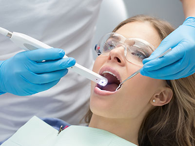
Using an intraoral camera is a straightforward, noninvasive part of a typical dental visit. The clinician or hygienist will guide the small camera into the mouth for a few moments while you relax in the chair. Bright, focused LED lighting and a steady hand produce crisp images; many patients notice only a mild awareness of the device, similar to the feeling of a mirror during an exam.
The images appear on the nearby monitor so you and your provider can view them together. Your clinician will point out areas of interest, explain findings in clear, understandable terms, and discuss any recommended next steps. If needed, still images are saved to your chart for future comparison or to share with other specialists and laboratories.
Because the process is quick and comfortable, intraoral imaging is an excellent option for routine checkups as well as problem-focused visits. It supports a conservative approach to care—spotting minor issues early and monitoring changes over time—while helping you stay informed about the condition of your smile.
In summary, intraoral cameras enhance diagnosis, strengthen communication, and integrate smoothly with modern dental technology to support precise, patient-centered care. If you’d like to learn more about how this imaging tool is used during routine exams or to assist with specific treatments, please contact us for more information.


An intraoral camera is a compact, pen-sized imaging device that captures high-resolution, full-color views of the teeth, gums and soft tissues. The camera uses focused LED lighting and close-range optics to reveal surface detail and texture at magnifications greater than the naked eye. Images transmit to a chairside monitor in real time so the clinician and patient can view the mouth together.
The device records still photos and short video clips that are stored with the patient record for documentation and comparison. Because the images are instantaneous, they support immediate discussion and help convert verbal findings into visible evidence. Many clinicians pair intraoral imagery with other diagnostic tools to create a complete clinical picture.
Dentists use intraoral cameras to improve visualization of subtle surface changes such as hairline cracks, early decay and localized tissue inflammation. Seeing magnified, lit images helps clinicians detect problems that might be difficult to notice with routine visual inspection alone. Real-time viewing also supports clearer communication between clinician and patient during the exam.
The images become part of the chart, so clinicians can track changes over time and document treatment needs. Visual records reduce ambiguity when coordinating care with other providers or laboratory technicians. Overall, intraoral cameras enhance diagnostic thoroughness and patient understanding without adding invasiveness to the visit.
No, an intraoral camera is a complementary tool rather than a replacement for X-rays or other radiographic imaging. X-rays reveal internal structures such as tooth roots, bone levels and interproximal decay that surface photography cannot show. Intraoral images focus on surface detail, color and texture to supplement the information gained from radiographs.
Clinicians choose the appropriate combination of imaging methods based on the diagnostic question and the patient's needs. When visual clues from a camera suggest deeper issues, radiographs or three-dimensional imaging may be used to refine the diagnosis. Using both surface and internal imaging provides a more complete view of oral health.
Yes, intraoral cameras are noninvasive and designed for patient comfort during routine exams. The wand-like device is small and lightweight, and many patients report only a mild awareness similar to holding a mirror in the mouth. LED illumination is safe and focused, providing clear images without causing discomfort.
Clinics follow strict infection control protocols such as disposable sleeves, barrier films and thorough sterilization of any reusable components. Clinicians are trained to position the camera gently and to accommodate patients who have sensitivity or a strong gag reflex. Overall, the procedure is quick and well tolerated by most patients.
High-quality intraoral photos provide objective visual evidence that supports diagnosis and treatment decisions. Images help clinicians evaluate restorative margins, tooth wear, soft-tissue conditions and shade relationships, which improves preparation and communication with dental laboratories. Clear visuals also allow clinicians to annotate areas of concern and explain recommended steps to patients in concrete terms.
When planning restorations, these images serve as a reference for contour, color and occlusal relationships, contributing to better-fitting, more esthetic outcomes. Saved photos create a baseline for monitoring healing or the longevity of restorations over multiple visits. This visual documentation streamlines case presentation and helps align expectations among all parties involved in care.
Yes, intraoral camera images can be shared with specialists, dental laboratories and other members of the care team to improve coordination. Digital files allow clinicians to provide precise visual references for shade matching, contour and specific areas of concern, which reduces misunderstanding and speeds collaborative planning. Most offices obtain patient consent before transmitting images to outside providers.
When sharing files, practices use secure transfer methods and follow privacy and data protection protocols. Standardized, well-lit images are easier for receiving clinicians and technicians to interpret, which supports consistent treatment outcomes. Patients who prefer to have copies of their images can request them through the office.
Intraoral images are typically linked to the patient s electronic health record and stored on secure office servers or cloud systems maintained by the practice. Images become a formal part of the chart and are organized so clinicians can retrieve them during subsequent visits for comparison or treatment planning. Regular backups are used to preserve clinical documentation and prevent data loss.
Access to stored images is restricted to authorized staff in accordance with the practice's privacy policies and applicable regulations. Offices maintain procedures for secure access, auditing and transmission of digital records. Patients can inquire about how their images are stored and request copies or further information from the practice team.
During an intraoral camera exam, the clinician or hygienist will briefly guide the small camera into the mouth while you recline in the chair. Images will appear instantly on a nearby monitor so you and the clinician can review the findings together. The procedure generally takes only a few moments and is comfortable for most people.
The clinician will point out areas of interest, explain what the images reveal and discuss any recommended next steps in clear terms. If appropriate, still photos are saved to your chart for future comparison or to share with other providers. Patients are encouraged to ask questions so they fully understand what the images show.
Intraoral imaging supports preventive care by making early signs of disease more visible, which can lead to less invasive interventions. Detecting small areas of enamel breakdown, localized inflammation or early restorative failure allows clinicians to intervene sooner and often preserve more natural tooth structure. Documented images also provide a visual record that motivates patients to follow recommended home-care strategies.
During routine checkups, clinicians use intraoral photos to compare changes over time and to track the effectiveness of preventive measures. This longitudinal perspective helps prioritize care and refine recall intervals for individual patients. By combining visual documentation with clinical assessment, practices can deliver more targeted preventive guidance.
Yes, Vaccaro Aesthetic and Family Dentistry incorporates intraoral imaging into routine exams and hygiene appointments to enhance communication and documentation. The practice uses the images to show patients what the clinician observes, to record current conditions and to support coordinated care when specialists or laboratory input is needed. Including visual documentation as part of standard visits helps make examinations more transparent and informative.
Patients at the practice can expect clinicians to explain images in plain language and to save relevant photos in the chart for future comparison. If an image suggests the need for additional diagnostic steps, the team will discuss possible next steps and answer questions to help patients make informed decisions. The use of intraoral cameras is one of several digital tools the practice employs to deliver thorough, patient-centered care.

Ready to book your next dental visit or learn more about our services?
Getting in touch with Vaccaro Aesthetic and Family Dentistry is quick and easy. Our friendly team is here to help with scheduling, answering questions about treatments, and addressing any concerns. Whether by phone or our convenient online form, we make connecting with us easy. Take the first step toward a healthier, more confident smile—contact us today and experience personalized dental care that truly makes a difference.

Back to top
