
A Panorex X-ray — often called a panoramic radiograph — captures a wide, two-dimensional image of the entire mouth in a single exposure. Unlike small intraoral images that focus on individual teeth, this extra-oral image displays all the teeth, the upper and lower jawbones, the temporomandibular joints (TMJ), and adjacent structures such as the nasal sinuses. That broad perspective makes it an essential first step in many diagnostic and treatment-planning scenarios.
Clinically, a Panorex is invaluable for spotting issues that might be missed on isolated films: unerupted or impacted teeth, developmental anomalies, large restorations or prostheses, jaw cysts and tumors, and signs of advanced periodontal bone loss. It also helps dentists evaluate tooth position and jaw relationships prior to more complex treatments such as implants, orthodontics, or reconstructive procedures. For patients, that means a clearer roadmap for care and fewer surprises down the line.
Because of its comprehensive nature, a Panorex is frequently used alongside other imaging modalities rather than as a replacement. It offers an efficient overview that guides the clinical team in deciding whether more targeted or three-dimensional imaging is necessary. At the practice level, this makes planning more precise, speeds up diagnosis, and supports coordinated care with specialists when needed.
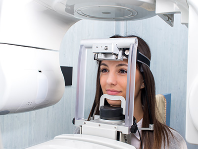
The Panorex exam is designed to be simple and comfortable. The patient stands or sits while placing the chin on a small rest and gently biting down on a plastic bite block to stabilize the jaw. During the exposure, the machine’s arm rotates around the head while the patient remains still for a matter of seconds. Because the image is taken from outside the mouth, there is no need for film or sensors to be placed between the teeth.
This noninvasive approach reduces discomfort and makes the procedure suitable for a wide range of patients, including those who have strong gag reflexes or difficulty tolerating intraoral sensors. Our team provides clear, step-by-step instructions and support throughout the process to ensure patient comfort and optimal image quality. If a patient has specific mobility or positioning needs, staff will adapt the procedure to accommodate them safely.
Once captured, the digital Panorex image is available immediately for review. The dentist can display the image on a computer screen to explain findings and discuss treatment options in real time. That visual context helps patients better understand the diagnosis and the rationale behind recommended next steps.
A Panorex image is particularly helpful for identifying impacted teeth, such as wisdom teeth that may be angled or trapped beneath the gumline. It also shows the presence and position of unerupted teeth in children and adolescents, which is useful for growth monitoring and orthodontic planning. In adults, a panoramic view can reveal extensive decay, large restorations, or fractures that affect multiple teeth or support structures.
Beyond teeth, Panorex films provide crucial information about the jawbone. Dentists use them to detect cysts, tumors, and abnormal bone patterns that require further evaluation. The TMJ area is also visible, allowing clinicians to spot changes that could relate to jaw pain or dysfunction. In addition, because the sinuses are partially included in the image, the Panorex can sometimes reveal signs of sinus-related issues that overlap with dental symptoms.
For periodontal assessment, the panoramic view helps visualize generalized bone loss and the overall architecture of the supporting jaw. While periapical or bitewing films give detailed views of individual teeth, the Panorex adds context by showing how periodontal changes relate across the entire dental arch — information that can influence comprehensive treatment planning.
Modern Panorex units produce high-quality digital images while keeping radiation exposure low. Digital sensors and updated imaging protocols require far less radiation than older film-based systems. When used appropriately, a panoramic radiograph provides a favorable balance between diagnostic yield and patient safety, particularly for screening and broad evaluations.
Radiation dose from a single Panorex is generally comparable to or lower than many other common medical imaging studies and is higher than a single intraoral bitewing but lower than full-mouth series comprised of multiple intraoral films. Dentists follow the principle of ALARA — “as low as reasonably achievable” — to minimize exposure, using panoramic films only when clinically justified and adapting settings for children or sensitive patients.
Pregnancy considerations are handled with care: routine dental imaging is typically deferred during pregnancy unless the information is essential for immediate care. When imaging is necessary, proper shielding and positioning are used to protect the patient. Our team reviews medical history and follows established safety guidelines before recommending any radiographic procedure.
Dentists commonly request a Panorex for initial evaluations, new-patient exams, oral surgery planning, and orthodontic assessments. It is particularly useful before wisdom tooth extractions, implant placement, and full-arch restorative work because it provides a comprehensive view of tooth relationships and the quality of available bone. Oral surgeons, orthodontists, and general dentists frequently use the Panorex as a baseline image that helps coordinate interdisciplinary care.
If the Panorex identifies abnormalities or areas of concern, it often prompts targeted follow-up imaging — for example, periapical x-rays for finer detail around a specific tooth or a cone beam CT (CBCT) scan for three-dimensional assessment of bone structure and nerve location. Using the panoramic film as a screening tool makes the diagnostic process more efficient and reduces unnecessary testing.
Throughout planning and treatment, the Panorex serves as a communication tool. It helps clinicians explain conditions and proposed procedures to patients, facilitates referrals when specialist input is needed, and documents the starting point for future comparisons. When integrated thoughtfully into care, a Panorex contributes to safer, more predictable outcomes and an improved patient experience.
In summary, the Panorex X-ray is a fast, comfortable, and informative imaging option that provides a broad view of teeth, jaws, TMJ structures, and neighboring anatomy. It supports diagnosis, treatment planning, and collaboration with specialists while prioritizing patient comfort and safety. If you have questions about whether a panoramic radiograph is appropriate for your needs, please contact Vaccaro Aesthetic and Family Dentistry for more information. Our team is happy to discuss how this imaging option may fit into your personalized care plan.


Panorex X-ray refers to a panoramic radiograph that captures a broad two-dimensional image of the entire mouth in a single exposure. The image includes all teeth, the upper and lower jawbones, the temporomandibular joints and portions of the nasal sinuses, providing a comprehensive overview of oral structures. As a screening tool, the Panorex helps clinicians identify areas that may need closer evaluation or targeted imaging.
The digital nature of modern panoramic units allows images to be available immediately for review on a computer screen. Dentists use the image to explain findings and outline possible next steps, which improves communication and helps patients understand recommended care. Because it shows the big picture, the Panorex often serves as the first step in diagnostic and treatment planning workflows.
A Panorex is an extraoral, or outside-the-mouth, image that displays the full dental arches and surrounding bone, while intraoral x-rays such as periapicals and bitewings focus closely on one or several teeth. That difference means intraoral films provide higher detail for individual tooth surfaces and root tips, whereas a panoramic view emphasizes overall relationships and large-scale abnormalities. Clinicians choose between these modalities based on whether they need fine local detail or a broader anatomical overview.
In practice, the two types of imaging are complementary rather than interchangeable. A Panorex can reveal areas of concern that prompt more focused intraoral films, and intraoral radiographs can confirm specific findings that are suggested on the panoramic view. Using both appropriately improves diagnostic accuracy and treatment planning efficiency.
A panoramic radiograph is especially useful for identifying impacted or unerupted teeth, large restorations, jaw fractures, cysts and tumors, and generalized periodontal bone loss across the arches. It also provides visibility of the TMJ region and parts of the sinuses, which can reveal issues that overlap with dental symptoms. Because the image covers the entire dental apparatus, it can expose patterns or abnormalities that isolated films might miss.
The Panorex is also valuable for growth monitoring and orthodontic evaluation, showing how teeth relate to one another and to the supporting bone. When the panoramic film suggests an abnormality, clinicians typically recommend follow-up imaging or referral for additional diagnostic work. As a result, the Panorex often serves as a central tool in comprehensive treatment planning.
Preparation for a panoramic radiograph is minimal and generally requires only removing jewelry, eyeglasses, removable dental appliances and hair accessories that could interfere with the image. Patients should inform the dental team about any medical conditions, recent surgeries or if they are pregnant so the staff can apply appropriate precautions. Wearing comfortable clothing and following staff instructions for positioning helps ensure a clear image.
Children and patients with mobility or positioning limitations should notify the office in advance so staff can make accommodations. The technologist or dental assistant will provide step-by-step guidance during the exam to maintain comfort and steady positioning. Clear communication about medical history and special needs allows the team to tailor the procedure safely.
Modern digital panoramic units produce high-quality images while keeping radiation exposure low compared with older film systems, and clinicians adhere to the ALARA principle to minimize dose. A single Panorex typically exposes a patient to less radiation than a full-mouth series of intraoral films while providing a much broader field of view. Settings are adjusted for patient size and sensitivity to reduce exposure further when appropriate.
When imaging is necessary for diagnosis or treatment planning, protective measures such as lead aprons and thyroid collars are used as indicated, and pregnancy considerations are handled conservatively. Dentists review medical history and weigh benefits and risks before recommending radiographs, ensuring that each image is clinically justified. If additional or alternative imaging is needed, the team will explain the reasons and safety practices involved.
The Panorex procedure is typically quick and noninvasive, with the actual exposure lasting only a few seconds while the machine rotates around the head. The patient stands or sits, places the chin on a rest and gently bites a plastic block to stabilize the jaw, and is asked to remain still during the rotation to avoid blurring. The short, outside-the-mouth technique is generally comfortable and well tolerated by most patients.
Because images are captured digitally, the Panorex is available immediately for review and discussion with the clinician. Staff will display the image on a screen to explain any notable findings and outline next steps in treatment or diagnosis. The efficiency of the process helps streamline appointments and improve patient understanding of oral health issues.
A panoramic radiograph is frequently used as an initial evaluation before procedures such as dental implant placement or wisdom tooth extractions because it provides an overview of tooth position and bone availability. The Panorex can reveal relationships between teeth and important anatomical structures like the mandibular canal, helping clinicians identify factors that influence surgical planning. While it does not replace three-dimensional imaging when detailed bone measurements are required, it is an essential first step in planning.
If the Panorex indicates concerns that require more precise assessment, clinicians will recommend targeted follow-up imaging such as periapical films or cone beam CT scans to map bone volume and nerve location. This staged approach allows for efficient screening and tailored use of advanced imaging resources. Vaccaro Aesthetic and Family Dentistry coordinates imaging choices to support safe, predictable surgical outcomes.
A Panorex often serves as a screening tool that may identify findings warranting three-dimensional evaluation, for example when the clinician needs detailed information about bone quality, tooth root proximity to nerves, or complex pathology. Cone beam CT (CBCT) provides volumetric data that clarifies depth and spatial relationships which a two-dimensional panoramic image cannot fully depict. The decision to proceed to CBCT is based on the clinical question and the need for precise measurements for surgical or implant planning.
Other situations that prompt further imaging include suspected lesions that require characterization, unclear tooth root anatomy, or preoperative mapping for complicated extractions. Using a Panorex first reduces unnecessary advanced imaging by highlighting when CBCT or additional intraoral films are truly indicated. The dental team will explain the rationale for any recommended follow-up studies and how they influence treatment choices.
Yes, children can have Panorex X-rays when clinically indicated, such as for evaluation of unerupted teeth, orthodontic assessment, or tracking dental development. Because pediatric patients are more sensitive to radiation, clinicians use adjusted exposure settings and shielding to minimize dose while still obtaining diagnostically useful images. The Panorex is often preferred for broad growth assessments because it avoids placing sensors inside a child’s mouth, which can be uncomfortable for some patients.
For routine localized concerns, smaller intraoral films may sometimes be more appropriate, and the dental team will choose the modality that provides the necessary detail with the least exposure. Staff members are trained to position and support children safely and to explain the process in age-appropriate terms. When imaging is recommended, parents can expect clear communication about why the radiograph is needed and how safety is maintained.
Panoramic radiographs are reviewed by the treating dentist and, when appropriate, by specialists such as oral surgeons or orthodontists who are involved in a patient’s care. The image serves as a diagnostic and documentation tool that helps clinicians identify conditions, determine the extent of pathology and plan interventions. Displaying the image during consultations aids in explaining findings and aligning treatment goals with the patient.
At Vaccaro Aesthetic and Family Dentistry, Panorex images are integrated into the patient record and used to develop coordinated, personalized care plans that may include further imaging or specialist referrals. The practice emphasizes clear communication about next steps and documents baseline images for comparison during follow-up. Patients receive an explanation of recommended actions and how the imaging supports safe, predictable treatment outcomes.

Ready to book your next dental visit or learn more about our services?
Getting in touch with Vaccaro Aesthetic and Family Dentistry is quick and easy. Our friendly team is here to help with scheduling, answering questions about treatments, and addressing any concerns. Whether by phone or our convenient online form, we make connecting with us easy. Take the first step toward a healthier, more confident smile—contact us today and experience personalized dental care that truly makes a difference.

Back to top
